What is renal pelvis obstruction?
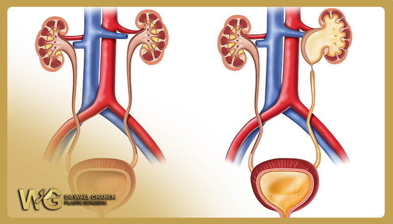
It is a blockage in the connection between the renal pelvis and the ureter, leading to enlargement of the renal pelvis, atrophy of kidney tissue, and a decrease in its efficiency. Typically, it affects only one kidney.
What are the causes of renal pelvis obstruction?
This obstruction is usually caused by underdeveloped nerves responsible for the expansion and contraction of the ureter in that area. Pressure on the junction of the ureter and renal pelvis may also be a contributing factor, often due to blood vessels.
What are the symptoms of renal pelvis obstruction?
In most cases, it is detected through ultrasound during pregnancy or diagnosed after birth when the following symptoms occur:
• Swelling on one side of the abdomen.
• Hematuria.
• Urinary tract infections.
• Kidney stones.
• Abdominal pain.
How is renal pelvis obstruction diagnosed?
The presence of swelling in the renal pelvis with narrowing at the beginning of the ureter is diagnosed through:
• Ultrasound imaging.
• Abdominal CT scan.
• X-ray with contrast material in the urinary tract.
• Renal scan.
What is the treatment for renal pelvis obstruction?
Firstly, the patient is closely monitored, and antibiotics are administered to prevent recurrent infections.
Secondly, if the renal pelvis is significantly enlarged, surgical intervention is performed to reconstruct the renal pelvis, widening the narrowed area. The procedure can be done laparoscopy. Postoperative follow-up is conducted to monitor kidney function.


